Enhanced in vitro mineralization and in vivo osteogenesis of composite scaffolds through controlled surface grafting of L-lactic acid oligomer on nano-hydroxyapatite.
作者:Wang, Zongliang; Xu, Yang; Wang, Yu ; Ito, Yoshihiro; Zhang, Peibiao*; Chen, Xuesi
关键字:Nanocomposite; poly(L-lactide); poly(lactide-co-glycolide); ceremic; scaffold; bone tissue engineering
论文来源:期刊
具体来源:Biomacromolecules,2016, 17 (3):818–829
发表时间:2016年
Abstract
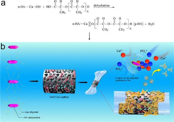
Nanocomposite of hydroxyapatite (HA) surface grafted with L-lactic acid oligomer (LAc oligomer) (op-HA) showed improved interface compatibility, mechanical property, and biocompatibility in our previous study. In this paper, composite scaffolds of op-HA with controlled grafting different amounts of LAc oligomer (1.1, 5.2, and 9.1 wt %) were fabricated and implanted to repair rabbit radius defects. The dispersion of op-HA nanoparticles was more uniform than n-HA in chloroform and nanocomposites scaffold. Calcium and phosphorus exposure, in vitro biomineralization ability, and cell proliferation were much higher in the op-HA1.1 wt %/PLGA scaffolds than the other groups. The osteodifferentiation and bone fusion in animal tests were significantly enhanced for op-HA 5.2 wt %/PLGA scaffolds. The results indicated that the grafted LAc oligomer of 5.2 or 9.1 wt %, which formed a barrier layer on the HA surface, prevented the exposure of nucleation sites. The shielded nucleation sites of op-HA particles (5.2 wt %) might be easily exposed as the grafted LAc oligomer was decomposed easily by enzyme systems in vivo. Findings from this study have revealed that grafting 1.1 wt % amount of LAc oligomer on hydroxyapatite could improve in vitro mineralization, and 5.2 wt % could promote in vivo osteogenesis capacity of composite scaffolds.
详见:http://pubs.acs.org/doi/abs/10.1021/acs.biomac.5b01543