生物体内承载组织,大多具有较强的生物力学性能,但是其弱的自修复能力导致其在发生损伤时,很难恢复到原来的结构和性能,组织工程替代物是实现承载组织功能再生的有效措施。水凝胶作为一种软湿材料,具有良好的生物相容性,在生物组织工程领域已经得到了广泛的应用,然而,由于其较低的力学性能(弱的抗张强度,较低的杨氏模量以及较差的韧性等),以及在水环境中不稳定的缺点,限制了其在生物承载组织中的应用。近年来,高强水凝胶获得了广泛而又深入的研究,并且取得了一定的成果,然而,由于高强水凝胶相对较低的模量,限制了其在跟腱、半月板等高模量生物承载组织的应用,而目前高模量水凝胶制备的复杂性、耗时性以及水环境中的不稳定性等缺点,使其作为生物组织替代物的研究更是鲜有报道。因此,设计一种简单有效地制备高强、高模量水凝胶,并能够在水中、PBS中保持稳定的水凝胶的方法仍然存在很大的挑战。
基于上述考虑,天津大学刘文广教授团队提出了一种力学“增强因子”的概念,通过一种全新的氢键单体NASC(只将已报道的NAGA侧链两个酰胺间的CH2换成NH,使NASC侧链带有一个酰胺键和一个脲基)的设计与合成,成功制备了PNASC超分子聚合物水凝胶。由于侧链强的氢键交联作用,溶胀平衡之后的超分子聚合物水凝胶的拉伸强度可达1.7-4.7 MPa,杨氏模量达48.4-100.3 MPa,断裂应变达341-638%,韧性可达5.66-20.35 MJ m-3,远高于迄今为止报道的超分子聚合物水凝胶 (Fig. 1),该团队对于PNASC水凝胶力学增强机制进行了探索。
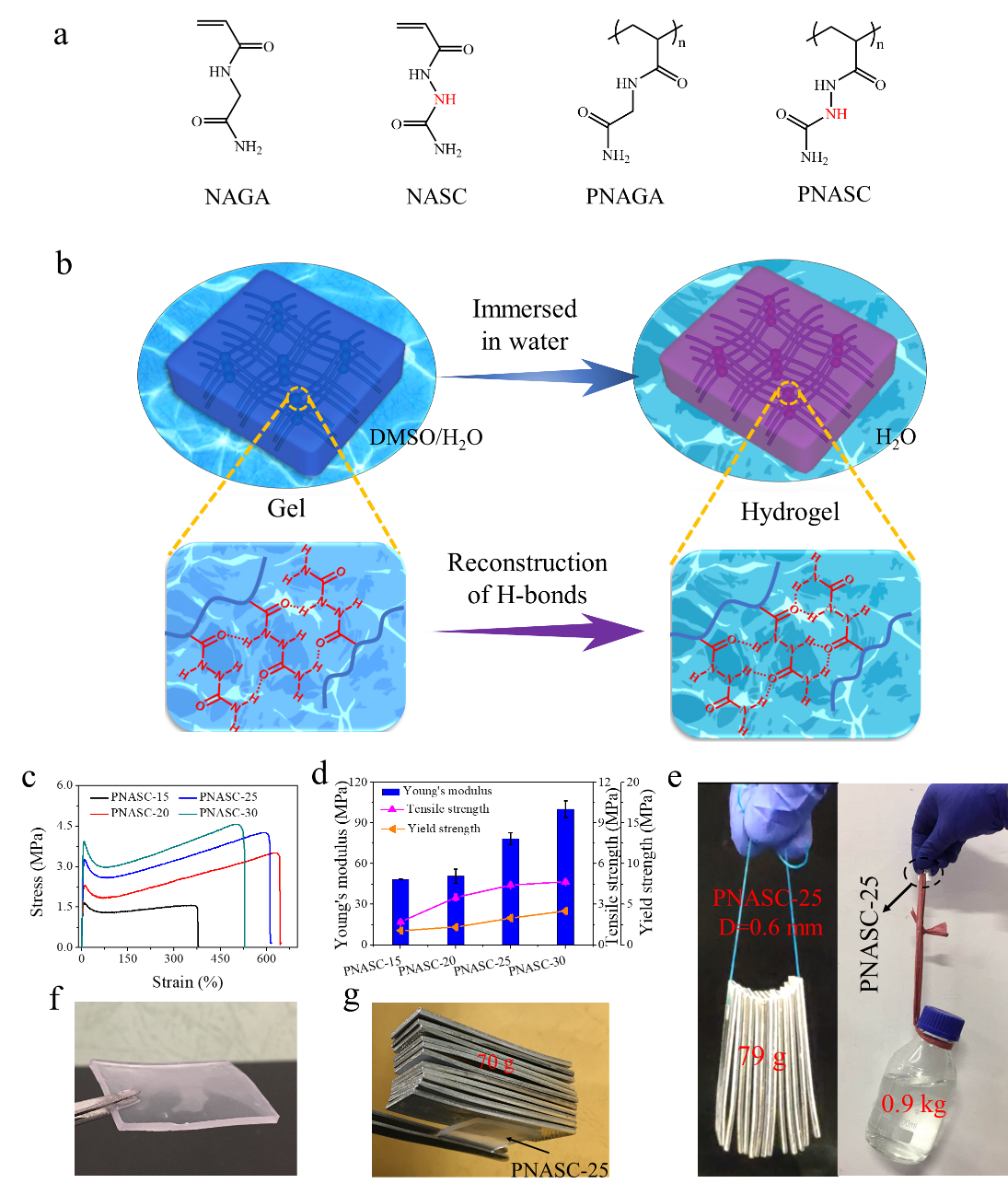
Fig. 1. (a): Molecular structures of NAGA, NASC, PNAGA and PNASC. (b): Schematic illustration of reconstruction of hydrogen bonding of amide and urea in PNASC gels when DMSO is replaced with water. (c): Tensile stress-strain curves. (d): Tensile strength, Young’s modulus and yielding strength of PNASC hydrogels with different initial monomer concentrations (n=3). (e, f, g) Photos portraying the high stiffness of the PNASC-25 hydrogels. (e): 79 g of aluminum sheets can be lifted up by a length of hydrogel line (d=0.6 mm) without stretching, and nylon cord is threaded through a hole in a piece of PNASC-25 hydrogel sheet (3 cm × 1.5 cm × 0.2 cm) and lifts a bottle of water weighing 0.9 kg without occurrence of deformation of the hydrogel hole. The hydrogel sheet (3 cm × 2.5 cm × 0.2 cm) was able to bear self-weight (f) and 70 g of aluminum sheets without collapse (g).
此外,值得注意的是,该单体可以与AAm,CBAA等代表性的单体共聚制备具有一定力学强度且在PBS中保持稳定的超分子聚合物水凝胶。为了探索PNASC基水凝胶的应用,该团队将单体NASC与接双键的肝素共聚,制备P(NASC-co-HepMAm) 共聚水凝胶微管,该水凝胶在37 ℃可以保持高的模量,并且具有良好的生物相容性和血液相容性,肝素的引入提高了其抗凝血性能,使其有望作为临时血管用于野外突发事故导致的血管破裂而引起的肢体缺血的救治,对于紧急情况下的受伤肢体的血液快速恢复具有潜在的应用价值 (Fig. 2)。
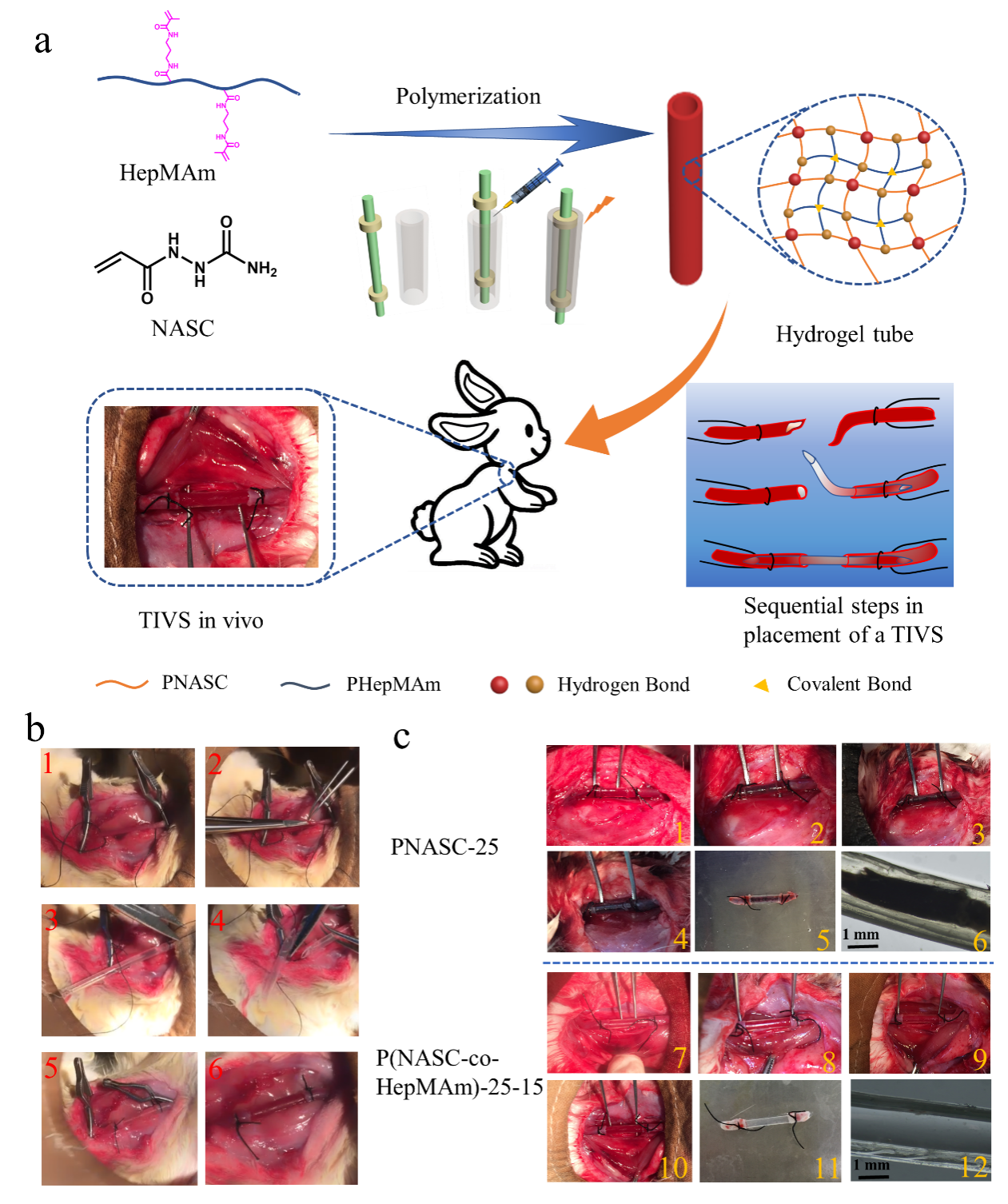
Fig. 2. (a) Schematic illustration of the fabrication of P(NASC-co-HepMAm) hydrogel tubes and their application as the TIVS in vivo. (b) Sequential steps in placement of hydrogel tube for TIVS in a rabbit model: (1) The rabbit’s arteria carotis was clamped on both sides by the artery clamp; (2) Defective treatment of blood vessels located between two arterial clips; (3) One end of the trimmed hydrogel tube was inserted into the rabbit’s carotid artery (3-4 mm depth) through the defect; (4, 5) Another end of the hydrogel tube was trimmed and the rabbit carotid artery was amputated; immediately, this end of the trimmed hydrogel tube was inserted into rabbit carotid artery (3-4 mm); (6) The arterial clip was removed to restore the blood flow. (c) (1, 7), (2, 8), (3, 9), and (4, 10) are the photos of blood color change in the PNASC-25 and P(NASC-co-HepMAm)-25-15 hydrogel tubes after being implanted for 0, 1, 2, and 4 h. (5, 11) are the photos of the PNASC-25 and P(NASC-co-HepMAm)-25-15 hydrogel tubes removed from the end of arteria carotis. (6, 12) Stereomicroscopic images of the PNASC-25 and P(NASC-co-HepMAm)-25-15 hydrogel tube luminal surface.
这项研究开辟了一种非常简单且通用的制备具有特定功能的高强度、高刚度、高韧性以及溶胀稳定的超分子聚合物水凝胶的策略,对于其在承载组织的应用中具有重要的意义。
目前上述工作已发表在《Materials Horizons》上,论文第一作者为天津大学材料学院博士生范川川,通讯作者为天津大学材料学院刘文广教授。
论文链接:https://pubs.rsc.org/en/content/articlelanding/2020/mh/c9mh01844a#!divAbstract
- 中国科大李家文教授团队 ACS Nano:3D打印源自单组分前驱体树脂的超高强度、超高韧性微纳SiOC陶瓷 2026-07-09
- 武汉大学常春雨教授课题组 AFM:高强度、各向异性矿化甲壳素可吸收螺钉 2026-06-04
- 吉林大学孙俊奇教授、李懿轩副教授团队 AFM: 基于二茂铁构象转变构筑低滞后、高强度与高韧性无液体离子导体 2026-06-02
- 清华大学徐军课题组 Macromolecules:非均匀交联构建高刚度、高韧性的形状记忆自愈合材料 2024-12-01
- 王双飞院士/聂双喜教授 Adv. Funct. Mater.:Hofmeister效应构筑摩擦电气凝胶 2023-09-04
- 新加坡科技设计大学Joel Yang 教授团队《Nano Lett.》:用于高分辨率可重构纳米光学的高刚度形状记忆聚合物 2022-11-15
- 安徽理工大学/都柏林大学王文新教授团队 AFM 封面:刚柔协同网络如何兼顾水凝胶的可注射性与高韧性 2026-06-26