开发满足不同特定需求的功能材料在生物医学中应用具有重大的挑战性。树枝状聚合物,凭借其精确的结构和多价协同性,是定制精密功能材料典范。近日,法国国家科学研究中心-马赛跨学科纳米研究中心与中国药科大学合作并成功开发了一种基于bola型两亲性树形分子(bola-amphiphilic dendrimers)的精确载体平台, 可选择性递送不同类型核酸药物(siRNA和DNA)(Figure 1),用于肿瘤治疗。结果发表在综合性期刊Proceedings of the National Academy of Sciences of the U. S. A.(DOI: 10.1073/pnas.2220787120)
合作团队设计和开发了不同代数的bola型两亲性树形分子并对其递送不同核酸分子的能力进行了研究。结果表明,bola树形分子的代数、核酸分子的大小以及bola树形分子与核酸分子之间多价协同作用在很大程度上影响其选择性递送核酸分子的能力。例如,更高代数的bola8A能更有效地将大尺寸质粒DNA 压缩成小的纳米复合物,显著提高了DNA在靶细胞的摄取,展现出更为优异的DNA转染活性;较低代数的bola4A则在有效保护短链siRNA分子的同时,能更高效地促进siRNA 的胞内释放,进而发挥强有力的基因沉默效果 (Figure 2)。此外, 该类bola两亲性树形分子所构建的核酸药物递送系统能选择性富集在肿瘤组织,并在肿瘤细胞内ROS刺激下特异性地释放核酸,实现核酸药物在肿瘤细胞的靶向递送。更令人欣喜的是,上述递送体系在宫颈癌和卵巢癌异种移植小鼠肿瘤模型、以及高侵袭性的黑色素瘤和三阴性乳腺癌转移模型中,均表现出可媲美商业化载体的核酸递送效率,能够精准调控致病基因的表达,发挥高效的抗肿瘤活性和抗肿瘤转移效果 (Figure 3)。本研究成果彰显了bola型两亲性树形分子作为按需定制核酸药物递送载体的巨大潜力。
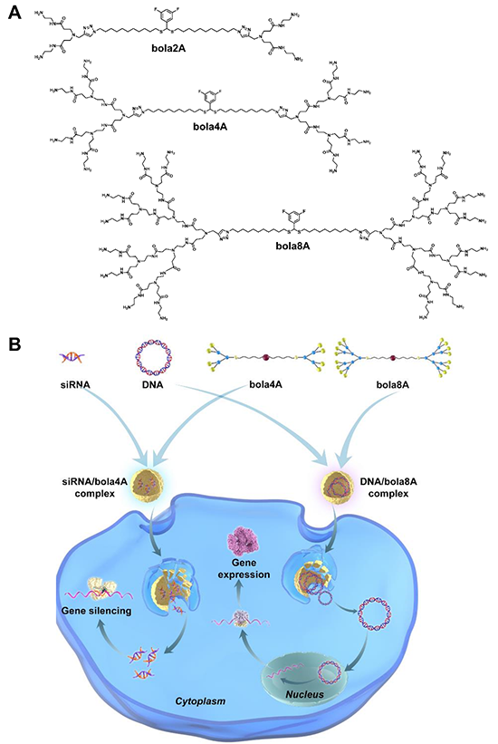
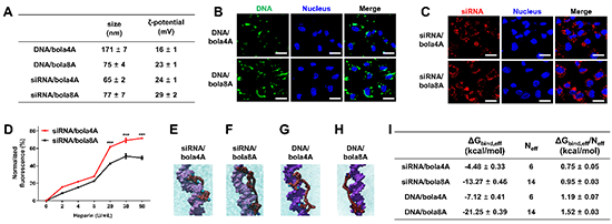
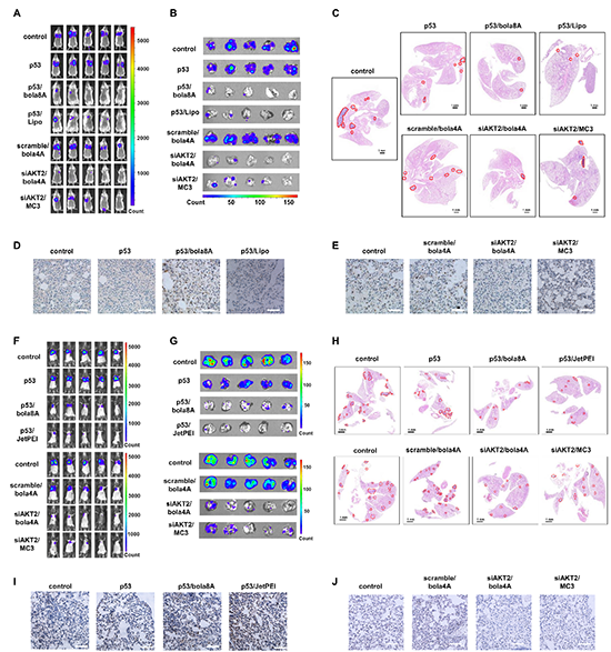
Fig. 3. Effective inhibition of tumor metastasis using DNA and siRNA therapeutics delivered by bola8A and bola4A respectively in lung metastatic cancer model. (A-E) 4T1-luc metastatic tumor-bearing mice received intravenous injections of PBS buffer (control), p53 alone, p53/bola8A complex, p53/Lipo complex (2.0 mg/kg DNA, 1.5 mg/kg bola8A, N/P ratio of 1.0), siAKT2 alone, siAKT2/bola4A complex, or siAKT2/MC3 complex (1.0 mg/kg siRNA, 3.9 mg/kg bola4A, N/P ratio of 5.0) (n=5): (A) In vivo bioluminescence imaging of 4T1-luc tumor metastases in the mice. (B) Ex vivo bioluminescence imaging of 4T1-luc tumor metastases in the lung at the experimental end point post treatment. (C) Histological analysis of lung tissues from 4T1-luc metastatic tumor-bearing mice at the experimental end point post treatment. The metastatic lesions (red solid outlines) were identified as cell clusters with darkly stained nuclei. Scale bars, 1 mm. (D) p53 and (E) AKT2 protein expression revealed by immunohistochemistry staining after treatments. Scale bar, 200 μm. (F-J) B16-F10-luc metastatic tumor-bearing mice received intravenous injections of PBS buffer (control), p53 alone, p53/bola8A complex, p53/JetPEI complex (2.0 mg/kg DNA, 1.5 mg/kg bola8A, N/P ratio of 1.0), siAKT2 alone, siAKT2/bola4A complex, or siAKT2/MC3 complex (1.0 mg/kg siRNA, 3.9 mg/kg bola4A, N/P ratio of 5.0) (n=5): (F) in vivo bioluminescence imaging of B16-F10-luc tumor metastases in the mice. (G) Ex vivo bioluminescence imaging of B16-F10-luc tumor metastases in the lung tissue or images of excised lung tissues at the experimental end point post treatment. (H) Histological analysis of lung tissues from B16-F10-luc metastatic tumor-bearing mice at the experimental end point post treatment. The metastatic lesions (red solid outline) were identified as cell clusters with darkly stained nuclei. Scale bars, 1.0 mm. (I) p53 and (J) AKT2 protein expression revealed by immunohistochemistry staining after treatments. Scale bar, 200 μm. p53: plasmid DNA expressing tumor suppressor protein p53, siAKT2: siRNA targeting AKT2.
原文链接:https://www.pnas.org/doi/10.1073/pnas.2220787120
- 法国艾克斯-马赛大学彭玲/中国药大刘潇璇/港大黄思齐 ACS AMI:两亲性树形分子包裹的伊马替尼针对转移性卵巢癌的有效治疗策略 2025-01-06
- 青岛大学于涛和亓洪昭团队《ACS AMI》:多阶段响应型纳米复合物通过改善口服核酸药物在结肠内的蓄积和分布来减轻溃疡性结肠炎 2022-04-08
- 天津大学仰大勇教授课题组《Nat. Commun.》:聚合物纳米框架中DNA时空编程级联组装赋能核酸药物精准递送 2021-02-19
- 中国科大王育才/蒋为团队 Nat. Nanotech.:首次揭示高分子纳米载体转移瘤递送新机制 2026-06-13
- 浙江大学申有青、周泉等 Nat. Commun.:DMSO万能溶剂启发的通用型高分子递药载体 2026-05-13
- 中科大王育才/朱书/蒋为团队 Science:系统揭示高分子载体材料发生系统性清除的底层机制 2026-03-20
- 哈工大冷劲松、哈医大二院原慧萍 AFM:智能药物递送低共熔凝胶用于青光眼长效治疗 2026-06-14